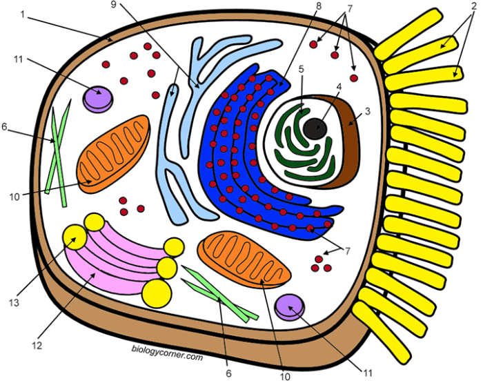
Illustrative Examples of Colored Animal Cells: Colored Animal Cell Coloring

Colored animal cell coloring – Microscopic examination of animal cells, following appropriate staining techniques, reveals a wealth of information about their structure and function. The choice of stain and the resulting coloration provide crucial visual cues for identifying different cell types and their subcellular components. Different staining methods highlight specific organelles or cellular structures, enabling detailed analysis of cellular morphology and physiology.
Stained Neuron Cell Appearance
A stained neuron, depending on the specific stain used, typically displays a characteristic morphology. The soma (cell body) often appears as a relatively large, round or irregularly shaped structure, stained a light to medium pink or purple with hematoxylin and eosin (H&E) staining. The nucleus, centrally located within the soma, stains a darker purple due to its high DNA concentration.
Extending from the soma are numerous dendrites, which are shorter, branching processes that appear as thinner, stained extensions, often exhibiting a similar pink or purple hue to the soma. The axon, a single, longer projection responsible for transmitting signals, is usually stained a lighter shade than the soma and dendrites, and may appear as a relatively straight, unbranched process.
Nissl bodies, regions of rough endoplasmic reticulum involved in protein synthesis, can be visible as basophilic (blue-purple) granules within the soma, particularly prominent with stains like cresyl violet. Myelin sheaths, if present, encasing the axon, may appear as a lighter, almost colorless region surrounding the axon, contrasting with the stained axonal process. The overall appearance is a complex network of stained processes emanating from a central, stained soma.
Stained Muscle Cell Appearance, Colored animal cell coloring
Stained skeletal muscle cells, or myofibers, typically exhibit a distinct striated appearance due to the highly organized arrangement of contractile proteins. Using H&E staining, the myofibrils appear as a series of alternating light and dark bands, reflecting the arrangement of actin and myosin filaments. The dark bands (A-bands) stain more intensely, usually a darker pink or purplish color, while the lighter bands (I-bands) appear a paler pink.
The nuclei of skeletal muscle cells are typically multiple, peripherally located, and elongated, staining a darker purple. Cardiac muscle cells, in contrast, exhibit branching patterns and intercalated discs, regions of specialized cell-to-cell junctions that appear as dark-staining lines between adjacent cells. Smooth muscle cells, however, appear spindle-shaped with a centrally located, elongated nucleus staining a darker purple, and lack the distinct striations of skeletal and cardiac muscle.
The cytoplasm of smooth muscle cells generally stains a lighter pink or pale purple. The variations in staining intensity and cellular arrangement allow for easy differentiation between these three types of muscle cells.
Stained Blood Cell Appearance
Stained blood cells reveal significant color variations depending on cell type and staining method. Using Wright-Giemsa stain, erythrocytes (red blood cells) appear as anucleated, biconcave discs that stain a characteristic bright pink or reddish-orange. Leukocytes (white blood cells) exhibit greater diversity in appearance. Neutrophils, the most abundant type, generally have a multi-lobed nucleus and a pale pink or light lilac cytoplasm with fine granules.
Lymphocytes typically have a large, round nucleus that occupies most of the cell, with a thin rim of pale blue cytoplasm. Monocytes display a larger, kidney-shaped nucleus and abundant light blue cytoplasm. Eosinophils are characterized by their large, reddish-orange granules, while basophils have dark purple granules that often obscure the nucleus. Platelets, small cell fragments involved in blood clotting, appear as small, irregularly shaped fragments staining light purple or pink.
The diverse coloration and morphology of blood cells enable their easy identification and differentiation under a microscope.
Visual Representation of Three Distinct Animal Cell Types
To visually represent three distinct animal cell types, consider a hypothetical microscopic field of view. A neuron could be depicted with a large, centrally located, dark purple nucleus within a pink soma, with numerous thinner, branching, pink dendrites extending outward. Adjacent to this, a skeletal muscle cell could be shown with its characteristic striations, alternating light pink and darker pink bands, and multiple peripherally located, dark purple nuclei.
Finally, a red blood cell, appearing as a small, anucleated, bright reddish-orange biconcave disc, could be interspersed among the neuron and muscle cell, highlighting the size and color differences between these three cell types. This illustrative representation emphasizes the distinct morphological and staining characteristics of these cells.
The Significance of Color in Animal Cell Biology

Cell color, while often overlooked, plays a crucial role in various aspects of animal cell biology. The inherent or acquired pigmentation of cells provides valuable insights into cellular function, intercellular interactions, and disease states. Understanding the mechanisms behind cellular coloration and its implications is therefore essential for advancing our knowledge of biological processes and developing effective diagnostic and therapeutic strategies.Cell coloration arises from the presence of various pigments within the cell, including porphyrins (like heme in hemoglobin), carotenoids, and melanins.
These pigments absorb and reflect specific wavelengths of light, resulting in the observed color. The distribution and concentration of these pigments are dynamically regulated, influenced by genetic factors, environmental stimuli, and cellular metabolic processes.
Cell Color and Cellular Function
The color of a cell can directly reflect its function. For example, the red color of erythrocytes (red blood cells) is due to the high concentration of hemoglobin, which is essential for oxygen transport. Similarly, the yellow-brown color of some cells in the liver is indicative of the presence of lipofuscin, a pigment associated with cellular aging and oxidative stress.
Variations in the intensity of these colors can signify changes in cellular activity or metabolic state. For instance, a decrease in the red color of erythrocytes could indicate anemia, while an increase in lipofuscin pigmentation might suggest cellular damage.
Cell Color and Disease States
Changes in cell color often serve as important diagnostic indicators of disease. For instance, the abnormal coloration of cells can be indicative of various pathologies. The yellowing of skin and sclera (jaundice) results from an accumulation of bilirubin, a byproduct of heme breakdown, indicating liver or blood disorders. Similarly, the presence of abnormal melanocyte activity (cells producing melanin) can lead to changes in skin pigmentation, sometimes associated with skin cancers like melanoma.
The aberrant coloration of cells observed in microscopic examination of tissue samples is routinely used in pathology to aid in disease diagnosis.
Future Research Directions in Cell Color Analysis
Future research could focus on developing more sensitive and specific methods for analyzing cellular coloration. Advanced imaging techniques, such as hyperspectral imaging, could provide detailed information on the spectral properties of cells, allowing for the identification and quantification of various pigments. This could lead to improved diagnostic tools for early disease detection and personalized medicine approaches. Furthermore, investigating the genetic and environmental factors that regulate cellular pigmentation could offer new therapeutic targets for treating diseases associated with abnormal cell coloration.
Examples of Advancements from Understanding Cell Color
The understanding of cell color has significantly contributed to advancements in various fields of biology. The study of hemoglobin’s role in oxygen transport, based on the red color of erythrocytes, has revolutionized our understanding of respiration and circulatory systems. Similarly, the study of melanin’s role in protecting against UV radiation, revealed through its dark pigmentation, has greatly enhanced our understanding of skin biology and the development of sunscreens and other protective measures.
The analysis of abnormal cell coloration in cytology and histology is integral to cancer diagnosis and treatment strategies.
Understanding the intricacies of colored animal cell coloring helps us appreciate the complexity of life. This detailed visualization is made easier when we see simplified representations, such as those found in animal coloring pages free printable. These pages offer a fun way to learn about animal anatomy, and subsequently, help visualize how the coloring in those pages can relate back to the actual colored components within a real animal cell.