Introduction to Animal Cells and Science Education: Science Is Real Animal Cell Coloring Page

Science is real animal cell coloring page – Early exposure to scientific concepts is crucial for fostering a lifelong appreciation of the natural world and developing essential critical thinking skills. A strong foundation in biology, beginning with fundamental cellular structures, provides a framework for understanding more complex biological processes and systems. Introducing children to the intricacies of animal cells at a young age lays the groundwork for future learning in various scientific fields, including medicine, genetics, and environmental science.Engaging visuals play a vital role in making abstract scientific concepts accessible and memorable for young learners.
Coloring pages, specifically those depicting animal cells with clearly labeled organelles, serve as an effective tool for visual learning. The act of coloring reinforces the names and locations of cellular components, transforming a potentially dry subject into an interactive and enjoyable experience. This visual reinforcement aids in memory retention and comprehension, bridging the gap between abstract knowledge and concrete understanding.Hands-on activities significantly enhance the learning process by transforming passive reception of information into active engagement.
The tactile nature of coloring, coupled with the opportunity to label organelles, creates a multi-sensory learning experience that promotes deeper understanding and knowledge retention. This active participation encourages critical thinking and problem-solving skills as children visually process the information and translate it into their coloring activity. The tangible aspect of the activity provides a lasting impression, making the learning process more meaningful and effective.
The Importance of Visual Aids in Cell Biology Education
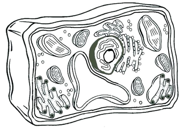
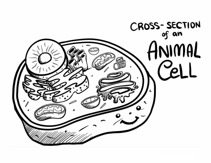
Visual aids, such as detailed diagrams and colorful illustrations of animal cells, significantly improve comprehension. For instance, a coloring page depicting a typical animal cell, with clearly labeled structures like the nucleus, mitochondria, and endoplasmic reticulum, allows students to visualize the spatial relationships between these organelles and understand their individual functions more effectively. The process of identifying and coloring each organelle reinforces their names and functions, making learning more interactive and less abstract.
The pedagogical value of a “science is real animal cell coloring page” extends beyond mere memorization; it fosters engagement with scientific concepts through visual representation. This hands-on approach contrasts sharply with the imaginative world presented in anime warrior coloring pages , highlighting the different cognitive processes involved. However, both activities share the common thread of creative expression and the development of fine motor skills, ultimately contributing to broader educational goals.
The visual representation aids in memorization and comprehension, surpassing the effectiveness of simply reading a textbook description. This method enhances the overall learning experience and allows for a more thorough understanding of animal cell structure and function.
Benefits of Hands-on Activities in Science Learning
Hands-on activities, such as coloring pages, provide a kinesthetic learning experience that improves engagement and knowledge retention. In the context of learning about animal cells, the act of coloring and labeling different organelles actively involves the learner, converting passive learning into an active, constructive process. This direct interaction enhances understanding and improves memory compared to passive learning methods like simply reading about the subject.
Furthermore, hands-on activities foster a sense of accomplishment and encourage exploration, stimulating curiosity and a deeper interest in the subject matter. The tangible nature of the activity allows for a more memorable and impactful learning experience, improving long-term retention of the information. For example, a student who colors and labels a cell diagram is more likely to remember the names and functions of the organelles compared to a student who only reads about them.
Animal Cell Structure and Function

Animal cells, the fundamental units of animal life, are complex structures containing numerous organelles, each with a specific role in maintaining cellular homeostasis and function. Understanding these components is crucial to grasping the intricate processes that govern life itself. This section details the structure and function of key animal cell organelles and explores the vital processes of cellular respiration and protein synthesis.
| Organelle | Function | Location within cell | Illustration Description |
|---|---|---|---|
| Cell Membrane | Regulates the passage of substances into and out of the cell; maintains cell shape and integrity. | Outer boundary of the cell. | A thin, continuous line surrounding the cell, often depicted as a flexible barrier with embedded proteins, resembling a mosaic. |
| Nucleus | Contains the cell’s genetic material (DNA), controlling cellular activities. | Usually centrally located, often the largest organelle. | A large, round structure containing a darker region (the nucleolus), depicted as a sphere with a speckled interior. |
| Cytoplasm | Gel-like substance filling the cell; site of many metabolic reactions. | Fills the space between the cell membrane and the nucleus. | A light gray or beige background filling the cell, providing a context for other organelles. |
| Mitochondria | Produce ATP (energy currency of the cell) through cellular respiration. | Scattered throughout the cytoplasm. | Small, oval structures with a folded inner membrane (cristae), often depicted as bean-shaped with internal lines representing the folds. |
| Ribosomes | Synthesize proteins based on genetic instructions. | Free-floating in the cytoplasm or attached to the endoplasmic reticulum. | Small, dark dots, either scattered or clustered along the endoplasmic reticulum. |
| Endoplasmic Reticulum (ER) | Network of membranes involved in protein and lipid synthesis and transport. Rough ER (RER) has ribosomes attached; smooth ER (SER) lacks ribosomes. | Extends throughout the cytoplasm. | A network of interconnected channels; RER is depicted with ribosomes attached, appearing rough; SER is depicted as smooth, continuous membranes. |
| Golgi Apparatus (Golgi Body) | Modifies, sorts, and packages proteins and lipids for secretion or transport within the cell. | Usually near the nucleus. | A stack of flattened sacs (cisternae), often depicted as a series of overlapping pancakes. |
| Lysosomes | Contain enzymes that break down waste materials and cellular debris. | Scattered throughout the cytoplasm. | Small, spherical vesicles containing darker contents, often depicted as small sacs with a dense interior. |
| Centrioles | Involved in cell division; organize microtubules. | Usually near the nucleus, often in pairs. | Small, cylindrical structures, usually depicted as paired structures near the nucleus. |
Cellular Respiration and Protein Synthesis
Cellular respiration, the process by which cells generate energy, occurs primarily within the mitochondria. The observable structures on a coloring page relating to this process are the mitochondria themselves. The folded inner membrane (cristae) significantly increases the surface area available for the metabolic reactions involved in ATP production. Protein synthesis, on the other hand, involves the coordinated action of ribosomes, the endoplasmic reticulum, and the Golgi apparatus.
Ribosomes, depicted as small dots, are the sites of protein synthesis. The endoplasmic reticulum, visualized as a network of channels, transports newly synthesized proteins to the Golgi apparatus, which further processes and packages them.
Analogy for Cellular Processes
Imagine a cell as a busy factory. The nucleus is the control center, containing the blueprints (DNA) for making products (proteins). Ribosomes are the assembly lines, building the products according to the blueprints. The endoplasmic reticulum is the conveyor belt, transporting the products to the packaging department (Golgi apparatus). Mitochondria are the power generators, providing the energy needed for the entire factory to operate.
Lysosomes are the waste disposal system, breaking down unwanted materials. The cell membrane is the factory’s security, controlling what enters and leaves.
Educational Activities using the Coloring Page
The animal cell coloring page serves as a versatile tool for engaging students in active learning and reinforcing their understanding of cell biology. Its effectiveness stems from its ability to cater to diverse learning styles, promoting both visual and kinesthetic engagement. The following activities leverage the coloring page to achieve a deeper understanding of animal cell structure and function.
The multifaceted nature of this learning tool allows for various pedagogical approaches, from independent study to collaborative projects, ultimately enhancing comprehension and retention.
Educational Activities for Diverse Learning Styles, Science is real animal cell coloring page
Utilizing the coloring page, educators can design activities to cater to the varied learning preferences within a classroom. This approach ensures inclusivity and maximizes the learning potential of each student. The following activities exemplify this approach:
- Visual Learners: Students can color-code organelles based on their function (e.g., all energy-producing organelles in one color, all membrane-bound organelles in another). This visual representation strengthens the connection between structure and function.
- Kinesthetic Learners: Students can create a three-dimensional model of an animal cell using the coloring page as a template. They can cut out the organelles and assemble them to create a tactile representation of the cell.
- Auditory Learners: Students can work in pairs, describing the function of each organelle to their partner while pointing to it on the coloring page. This verbalization process solidifies their understanding.
- Read/Write Learners: Students can label each organelle on the coloring page and write a brief description of its function. This combines visual learning with written expression.
Lesson Plan Incorporating the Animal Cell Coloring Page
This lesson plan integrates the coloring page to provide a structured learning experience about animal cell structure and function. The plan is designed to be adaptable to various grade levels with adjustments in complexity and detail.
- Introduction (10 minutes): Begin with a brief overview of cells as the basic units of life. Introduce the concept of animal cells and their key components.
- Activity 1: Coloring and Labeling (20 minutes): Distribute the coloring page. Students color and label the major organelles (nucleus, cytoplasm, mitochondria, ribosomes, endoplasmic reticulum, Golgi apparatus, lysosomes, cell membrane).
- Activity 2: Function Discussion (15 minutes): Engage students in a class discussion about the function of each organelle. Use visual aids and analogies to enhance understanding. For example, compare the mitochondria to a power plant, the Golgi apparatus to a post office, and the lysosomes to a recycling center.
- Activity 3: Group Work and Presentation (15 minutes): Divide students into small groups. Each group focuses on one or two organelles, researching their specific function in more detail and preparing a short presentation to the class.
- Assessment (10 minutes): A short quiz on the major organelles and their functions can be administered. The completed and labeled coloring pages can also serve as part of the assessment.
Assessing Student Understanding using the Coloring Page
The coloring page itself provides a valuable assessment tool. The accuracy of labeling, the completeness of coloring, and the overall presentation can offer insights into student comprehension. Further, specific questions can be posed based on the colored and labeled page to gauge deeper understanding.
- Accuracy of Labeling: Correctly identifying and labeling all major organelles demonstrates a solid understanding of animal cell structure.
- Color-Coding and Organization: A well-organized and color-coded page reflects a structured understanding of the relationships between different organelles and their functions.
- Written Descriptions (Optional): Requiring students to write short descriptions of each organelle’s function provides a more in-depth assessment of their knowledge.
- Follow-up Questions: Questions such as “How does the structure of the mitochondria relate to its function?” or “What would happen if the cell membrane were damaged?” can be used to assess higher-order thinking skills.