Introduction to Animal Cell Structure

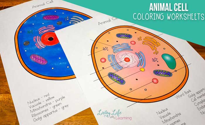
Animal cells coloring worksheet – Animal cells are the fundamental building blocks of animal tissues and organs. Understanding their structure is key to grasping how animals function at a cellular level. These cells, unlike plant cells, lack a cell wall and chloroplasts, resulting in a different overall structure and function. Let’s explore the major components.
The oddly satisfying task of coloring animal cells felt strangely familiar, a microscopic echo of something larger. The vibrant hues reminded me of the ocean’s depths, and I wondered if those same colors could be found in the intricate details of printable sea animals coloring pages. Perhaps the cellular structures, when magnified, held a secret key to understanding the vibrant life teeming within those creatures, mirroring the complexity of the animal cells coloring worksheet itself.
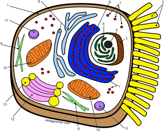
Animal cells are complex structures containing a variety of organelles, each with a specific role in maintaining the cell’s life and function. These organelles work together in a coordinated manner, ensuring the cell can carry out its essential processes, from energy production to protein synthesis. The key components we will focus on are the cell membrane, cytoplasm, nucleus, mitochondria, ribosomes, and endoplasmic reticulum.
Cell Membrane
The cell membrane, also known as the plasma membrane, is a selectively permeable barrier that surrounds the cell, separating its internal environment from the external environment. It’s composed primarily of a phospholipid bilayer, with embedded proteins that act as channels, transporters, and receptors. This structure allows the cell to regulate the passage of substances into and out of the cell, maintaining homeostasis.
The membrane’s fluidity allows for flexibility and movement, crucial for cell processes like cell division and signaling.
Cytoplasm
The cytoplasm is the jelly-like substance that fills the cell, excluding the nucleus. It’s a complex mixture of water, salts, and various organic molecules, including enzymes and proteins. The cytoplasm is the site of many metabolic reactions and acts as a medium for transport of materials within the cell. Organelles are suspended within the cytoplasm, and the cytoskeleton, a network of protein filaments, provides structural support and facilitates movement within the cell.
Nucleus
The nucleus is the control center of the cell, containing the cell’s genetic material (DNA). It’s enclosed by a double membrane called the nuclear envelope, which has pores that regulate the passage of molecules between the nucleus and the cytoplasm. Inside the nucleus, DNA is organized into chromosomes, which carry the genetic instructions for the cell’s functions. The nucleolus, a dense region within the nucleus, is involved in the synthesis of ribosomes.
Mitochondria
Mitochondria are often referred to as the “powerhouses” of the cell because they are responsible for cellular respiration, the process of converting nutrients into energy in the form of ATP (adenosine triphosphate). They have a double membrane structure, with the inner membrane folded into cristae, which increase the surface area for ATP production. Mitochondria also play a role in other cellular processes, including calcium storage and apoptosis (programmed cell death).
Ribosomes, Animal cells coloring worksheet
Ribosomes are small, granular organelles responsible for protein synthesis. They are composed of RNA and protein and can be found free in the cytoplasm or attached to the endoplasmic reticulum. Ribosomes translate the genetic information encoded in mRNA (messenger RNA) into proteins, which carry out a wide range of functions within the cell.
Endoplasmic Reticulum
The endoplasmic reticulum (ER) is a network of interconnected membranes extending throughout the cytoplasm. There are two types of ER: rough ER and smooth ER. Rough ER is studded with ribosomes and is involved in protein synthesis and modification. Smooth ER lacks ribosomes and plays a role in lipid synthesis, detoxification, and calcium storage.
| Cell Component | Function | Description | Illustration |
|---|---|---|---|
| Cell Membrane | Regulates passage of substances; maintains cell shape | A selectively permeable phospholipid bilayer with embedded proteins. | A double layered oval shape with various smaller shapes (proteins) embedded within. The outer layer and inner layer are slightly different shades, representing the phospholipid bilayer. |
| Cytoplasm | Site of metabolic reactions; transports materials | A jelly-like substance filling the cell, containing organelles and cytoskeleton. | A light-grey, gel-like background with various colored shapes (organelles) scattered throughout. Thin, interwoven lines represent the cytoskeleton. |
| Nucleus | Houses genetic material; controls cell activities | A spherical organelle enclosed by a double membrane (nuclear envelope) containing chromosomes and the nucleolus. | A large, dark-colored circle (nucleus) with a smaller, slightly lighter circle inside (nucleolus). A thin, double line represents the nuclear envelope. |
| Mitochondria | Cellular respiration; ATP production | Bean-shaped organelles with a double membrane; inner membrane folded into cristae. | Several bean-shaped organelles, each with a darker inner membrane folded into wavy lines (cristae). |
| Ribosomes | Protein synthesis | Small, granular organelles composed of RNA and protein. | Small, dark dots scattered throughout the cytoplasm and attached to the endoplasmic reticulum. |
| Endoplasmic Reticulum | Protein and lipid synthesis; detoxification | A network of interconnected membranes; rough ER has ribosomes, smooth ER does not. | A network of interconnected, thin, light-grey lines. Some areas are studded with small dark dots (ribosomes) representing rough ER, while others are smooth. |
Designing the Worksheet
Okay, so we’ve covered the basics of animal cell structure. Now let’s get this coloring worksheet looking awesome and informative. We want something engaging for students, not just a boring diagram. Think vibrant colors and a layout that makes learning fun.The key is simplicity. We don’t need to include every single organelle; we want a clean, easily understandable representation of the major components.
Overloading the diagram with detail will just confuse things.
Cell Structure Representation
The worksheet will feature a simplified, large-scale diagram of an animal cell. This allows ample space for coloring and labeling. The overall shape will be a slightly irregular circle, reflecting the typical animal cell morphology. The cell’s components will be clearly delineated and proportionally sized to each other, although not to scale in terms of real cellular dimensions.
Specific Structures Included
The following structures will be included in the diagram:
- Cell Membrane: This will be represented as a thin, outer boundary surrounding the entire cell. It’s the cell’s protective barrier.
- Nucleus: A large, centrally located circle representing the control center of the cell. It will be clearly differentiated from the cytoplasm.
- Cytoplasm: The jelly-like substance filling the cell, shown as the area between the cell membrane and the nucleus. It houses the organelles.
- Mitochondria: Several smaller oval shapes scattered within the cytoplasm. These are the “powerhouses” of the cell, generating energy.
Color Scheme and Rationale
Color choice is crucial for both visual appeal and clarity. We’ll use a scheme that helps students easily distinguish between the different cell parts.
- Cell Membrane: A light, pastel blue. This represents the fluid and permeable nature of the membrane.
- Nucleus: A deep, rich purple. This stands out and emphasizes the nucleus’s importance as the cell’s control center.
- Cytoplasm: A pale, light yellow. This provides a neutral background to highlight other organelles.
- Mitochondria: A bright, energetic orange. This represents the energy-producing function of the mitochondria. We could also consider a reddish hue for an alternative look.
These colors are chosen for their contrast and visual appeal, minimizing confusion and maximizing the learning experience. They also evoke a sense of life and activity within the cell.
Worksheet Activities and Extensions: Animal Cells Coloring Worksheet

To make learning about animal cells more engaging and cater to diverse learning styles, we’ve developed some fun activities to complement the coloring worksheet. These activities aim to solidify understanding through hands-on experience and different approaches to information processing. They also provide opportunities for assessment and extension beyond basic coloring.These activities are designed to reinforce the concepts learned through the coloring exercise, providing students with multiple avenues for understanding animal cell structure and function.
The activities offer a variety of approaches, from kinesthetic learning to visual reinforcement and critical thinking.
Three Engaging Activities
The following activities provide diverse learning experiences to complement the coloring worksheet. Each activity targets a different learning style, ensuring a more comprehensive understanding for all students.
- Build-a-Cell: Students use modeling clay or other craft materials to create a three-dimensional model of an animal cell. This kinesthetic activity allows students to physically interact with the cell structure, reinforcing their understanding of the relative sizes and positions of organelles. They should label each organelle using toothpicks and small flags or labels. This activity encourages collaboration and problem-solving as students work together to accurately represent the cell’s components.
- Cell Structure Bingo: Create bingo cards with images or names of different animal cell organelles. Call out the names or show images of organelles, and students mark them on their cards. This visually-oriented activity reinforces vocabulary and recognition of cell structures. The first student to get bingo wins a small prize. This game is a fun and competitive way to review the material.
- Animal Cell Comic Strip: Students create a short comic strip depicting the activities and interactions of different organelles within an animal cell. This creative writing activity encourages students to think about the functions of organelles in a narrative context. The comic strip can be used as a form of assessment, demonstrating understanding of the organelles and their roles within the cell. Students can be encouraged to be creative and humorous in their approach.
Multiple-Choice Questions
These multiple-choice questions assess understanding of key animal cell structures and their functions. Correct answers are crucial for demonstrating a grasp of fundamental cell biology concepts. These questions can be used as a quiz or review activity.
- The control center of the animal cell, containing the genetic material, is the:
- Ribosome
- Nucleus
- Mitochondrion
- Golgi Apparatus
(Correct answer: b)
- Which organelle is responsible for generating energy for the cell?
- Lysosome
- Endoplasmic Reticulum
- Mitochondrion
- Vacuole
(Correct answer: c)
- The packaging and distribution center of the cell is the:
- Cell Membrane
- Golgi Apparatus
- Cytoplasm
- Nucleolus
(Correct answer: b)
- Which organelle helps in protein synthesis?
- Centrioles
- Ribosomes
- Vacuoles
- Chloroplasts
(Correct answer: b)
- The jelly-like substance filling the cell is called:
- Cell Wall
- Cytoplasm
- Nucleoplasm
- Vacuole
(Correct answer: b)